OXALIA · Medical AI
AI doesn't replace the radiologist. It gives them back their time.
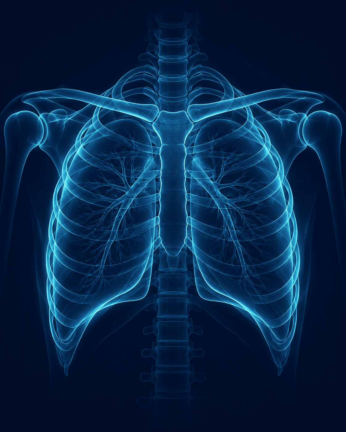
The first medical AI platform for chest radiography developed in Tunisia. Analysis in under two minutes, integrated with your PACS.
- Analysis
- < 2 min
- Pathologies
- 14
- Integration
- PACS/
DICOM - Institutional partners
- 3
Our institutional partners
01 · Our conviction
A medical desert that keeps growing.
Demand for medical imaging is growing faster than the number of practicing radiologists. Wait times lengthen, workload increases, and fatigue raises the risk of error on complex cases.
4
Radiologists (Tunisia)
13
Radiologists (ref.)
Radiology coverage by governorate
02 · Our response
A platform built to fit in, not to take over.
OXALIA fits into the existing workflow, with no new interface to learn, and a deployment model adapted to the realities of the Tunisian field.
Fast analysis
A structured result in under two minutes, ready to include in the report.
PACS/DICOM integration
Compatible with existing systems. No additional interface to learn.
Expert second opinion
Helps confirm a doubt or prioritize urgent cases — never a replacement for clinical judgment.
Built for the field
Cloud or on-premise, including over unstable connections or in mobile units.
03 · In the real workflow
The product in the real workflow, not in isolation.
Every exam lands in a prioritized worklist. One click opens the preliminary report generated by OXALIA, ready for the radiologist to validate.
Preliminary report
- Exam
- EXM-2024-1847
- Technique
- Frontal chest radiograph
Findings
Diagnostic aid — the final decision rests with the radiologist.
4 exams analyzed · 1 urgent cases prioritized
04 · What OXALIA detects
Every abnormality, located and named.
Hover or select a term to see how OXALIA locates it on the image.
OXALIA analyzes every chest radiograph and flags , , , , and — located, in under two minutes.
Cardiovascular
Air
Fluid
Mass
Parenchyma
Infectious
Skeletal
Reference
Diagnostic aid — the final decision rests with the radiologist.
See all detected pathologies05 · Evidence & method
Measured, published, compared.
OXALIA's performance is assessed against a public, comparable, and verifiable methodology — not a marketing promise.
Sensitivity
—
Specificity
—
Productivity gain
—
Regulatory status
OXALIA is not currently CE-marked or FDA-cleared. The regulatory path is published as soon as it is confirmed, rather than hidden.
06 · Ecosystem
A multidisciplinary team, locally rooted.
OXALIA is built by a team combining medical expertise, AI engineering, and regulatory compliance, working directly with Tunisian health institutions.
Radiology, machine learning, and regulatory affairs — team profiles will be published with their consent.
Ministère de la Santé
Scientific agreement
Hôpital Abderrahmen Mami
Partner clinical site
SUP'COM
Research laboratory
GET Wireless
Scientific agreement
07 · Roadmap
Four phases, forty-eight months.
OXALIA's development follows a progressive path, from the current detection module to automated reporting.
- Phase 1 — TODO
1. Chest detection
Rollout of the 2D detection module, initial clinical validation at a pilot site.
- Phase 2 — TODO
2. Regulatory expansion
Building the CE-marking file, expanding the number of pathologies covered.
- Phase 3 — TODO
3. 3D reconstruction
Launch of the 3D reconstruction module, expansion into other regional markets.
- Phase 4 — TODO
4. Automated reporting
Automated report generation and large-scale rollout, including mobile units.